What is a dental X-ray?
Dental X-rays, also known as radiographs, are images of your teeth, gums, and skeletal bones using low levels of radiation (X-rays) from a special machine.
Why is a dental X-ray necessary?
Not everything can be seen by looking into your mouth with a light and mirror. Most problems start in areas that are difficult or impossible to see, underneath the gums or in between teeth.
Dental X-rays are used to identify and treat dental problems such as cavities, infections, bone loss, fractured teeth, and the presence of foreign bodies.
Here are some common types of X-ray images taken in the dental office:
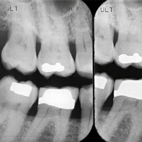
- Bitewing. These are pictures taken of the molars and premolars showing part of the upper teeth and part of the lower teeth together. Bitewings are the most efficient method for detecting cavities between the teeth as well as tooth decay under existing restorations.

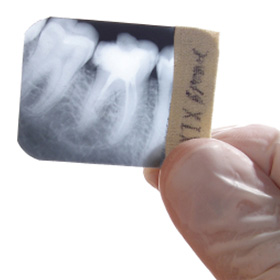
- Periapical. These are pictures taken of the whole tooth and bone around it. These are used to assess pain associated with a tooth, bone loss caused by gum disease, or root canal problems.

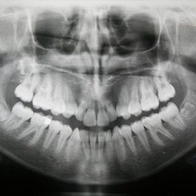
- Panoramic. This is a larger picture that shows the jaw bone, all the teeth in the mouth, and some boney structures in the head. These are useful for evaluating the development of teeth, bone fractures in or around the mouth area, detecting dental problems, and planning treatment such as orthodontics and implant placements.

Your dentist uses X-rays to identify dental problems, which allows for early treatment and proper planning before potential problems get more complicated and difficult to treat.
All material copyright MediResource Inc. 1996 – 2023. Terms and conditions of use. The contents herein are for informational purposes only. Always seek the advice of your physician or other qualified health provider with any questions you may have regarding a medical condition. Source: www.medbroadcast.com/healthfeature/gethealthfeature/Dental-Procedures
